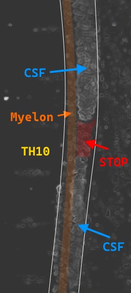
MR CSF flow study
As a supplement to the Facebook post describing the non-invasive, preoperative findings from 1 April 2009 (step 5 from the patient collective diagram), with images that were unique in the world at the time, and showing parts of an #aesiolysis operation on 2 April 2009.
A second diagnostic in this programme had previously taken place on 27 February 2009 and included a #CSF flow study (step 2).
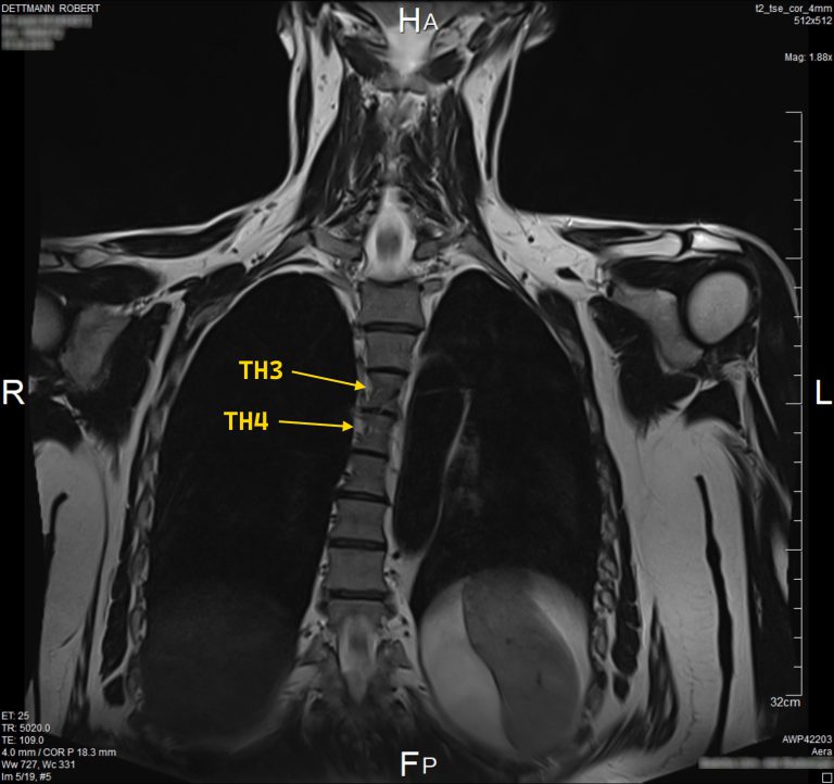
In that finding, a stop was detected at the level of BWK 10.
However, in the later, even more precise diagnosis of 1 April 2009, a further valve-like stop was seen at the level of BWK 3/ 4, where the maximum adhesion was assumed, although the findings of 27 February 2009 were initially more dominant.
During the adhesiolysis on 2 April 2009 at the level of BWK 3/4, however, the complex network of hyperplastic, arachnoid membranes running across the entire thoracic spine (finding from 1944) gradually collapsed.
This also solved the problem at the level of BWK 10.
The membranes are only severed and the remnants remain in the dural tube of the spinal canal.
This should help to prevent scarring and thus new adhesions.
Here you can also see a cine sequence of the CSF flow study at the level of BWK 10 and a section of this pulse-dependent movement of the CSF that I have reworked …