Arachnoid membrane hyperplasia
From 2001 onwards, due to the desolate healthcare system, I was once again on my own in search of my suspicion, which was only to be confirmed seven years later.
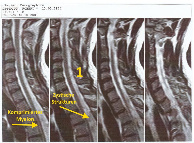
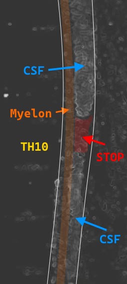
This suspicion concerned cystic changes in the spinal cord compressing the myelon and blocking the CSF, which none of the dozens of radiologists wanted to see.
Later it turned out that this would have been a top 100 diagnosis in neuroradiology.
Due to the misunderstanding of this diagnosis, I came up with the new, exact name “𝗣𝗼𝘀𝘁𝗶𝗻𝗳𝗲𝗸𝘁𝗶ö𝘀𝗲 𝗮𝗿𝗮𝗰𝗵𝗻𝗼𝗶𝗱𝗮𝗹𝗲 𝗠𝗲𝗺𝗯𝗿𝗮𝗻-𝗛𝘆𝗽𝗲𝗿𝗽𝗹𝗮𝘀𝗶𝗲”, which is based on almost 100-year-old findings on spinal and intracranial arachnoiditis.
This information was already available to me in 2004.
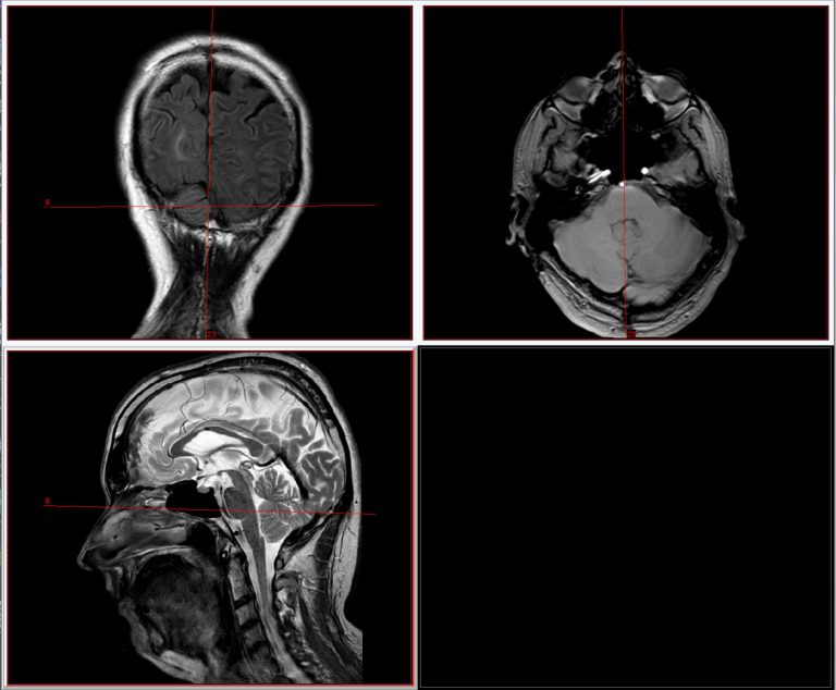
This was confirmed in 2009 as part of a research project in a newly developed worldwide diagnostic imaging method with intraoperative comparison.
In the video, I show in this newly developed dynamic BFFE cine sequence these now finally preoperatively visible, myelon-compressing, CSF-blocking, post-infectious multiplied membranes based on the spinal canal I can see there.
However, all of this was already known in the 1940s in the distant past, when it was found intraoperatively in all parts of the CNS in post-infectious patients.
Such a condition was known at that time even after banal colds.
This is shown by a paper published in 𝟭𝟵𝟰𝟰𝟰 by the University Hospital Zurich.
This is how it read for the findings of the histological examination from 1944:
“The findings of excised arachnoid pieces are variable.
Frequently, both intracranial and spinal forms of arachnoiditis are characterised by a strong
𝙃𝙮𝙥𝙚𝙧𝙥𝙡𝙖𝙨𝙞𝙚 𝙙𝙚𝙧 𝙖𝙧𝙖𝙘𝙝𝙣𝙤𝙞𝙙𝙚𝙖𝙡𝙚𝙣 𝙈𝙚𝙢𝙗𝙧𝙖𝙣𝙚𝙣.
Instead of loose connective tissue, they consist of a coarse network of thick collagen fibres.
The arachnoid epithelium, the covering cell layer, is frequently thickened, often five to seven layers thick.
The excised pieces of tissue usually lack inflammatory phenomena; sometimes only sparse fibrocytes are present.
Sometimes accumulations of round cells are found (case 8, 9, 15), occasionally also of epithelioid cells.
The thickened arachnoid membrane is here and there permeated by newly formed, delicate-walled vessels; the normal arachnoid membrane is without vessels (cases 11, 12, 13, 14).” …